


Newsroom

Advanced Imaging Technology for High-Resolution Analysis of Human Brain Hemispheres

For decades, the neuroscience community has aspired to observe the human brain in its entirety, both at large and microscopic scales. A recent study published in Science by a team based at MIT has introduced a groundbreaking technology pipeline that allows for the detailed processing, labeling, and imaging of complete brain hemispheres from two donors—one diagnosed with Alzheimer’s disease and the other without—achieving high resolution and speed.
Innovative Multi-Scale Brain Imaging Pipeline
Kwanghun Chung, the senior author and associate professor in the MIT departments of Chemical Engineering and Brain and Cognitive Sciences, highlighted the importance of this technology: “We performed holistic imaging of human brain tissues at multiple resolutions, from single synapses to whole brain hemispheres, and we have made that data available. This technology enables us to analyze the human brain at various scales, paving the way for comprehensive mapping of human brains.”
While this study does not deliver a complete atlas of the brain identifying every cell and protein, it showcases an innovative integration of three distinct technologies that facilitate extensive neuroscience research. The research serves as a proof of concept, revealing landscapes of thousands of neurons across various brain regions and offering detailed views of individual cells and subcellular structures. Additionally, the team provides quantitative analyses comparing regions within Alzheimer’s and non-Alzheimer’s hemispheres, highlighting the capabilities of their new approach.
Preserving Tissue Integrity for Comprehensive Analysis
The ability to image entire hemispheres intact at the resolution of individual synapses is crucial for understanding human brain health and disease. This technology allows scientists to investigate questions using the same brain sample, eliminating discrepancies caused by variations in different brains. A key advantage is that the analysis process preserves tissue integrity, making it more durable and re-labelable for future studies. In their work, Chung’s team utilized 20 different antibody labels to distinguish various cells and proteins, with plans to expand this to over a hundred labels.
Chung explained, “We need to visualize all functional components—cells, their morphology, connectivity, subcellular architectures, and individual synaptic connections—ideally within the same brain, given the variability present in human samples. This technology pipeline allows us to extract critical features from a single brain comprehensively.”
“This technology pipeline allows us to extract critical features from a single brain comprehensively.”
Advanced Tools Driving the Breakthrough
Moreover, the scalability and efficiency of this pipeline enable the creation of diverse samples that can represent various demographics, including sex, age, and disease states, thus facilitating robust comparative studies with increased statistical power. Chung envisions establishing a brain bank comprising fully imaged brains for researchers to analyze and re-label as required for new studies.
The project was made possible by a talented team at MIT, which included three co-lead authors responsible for three significant innovations. Ji Wang developed the “Megatome,” a device designed to slice intact human brain hemispheres without causing damage. Juhyuk Park created a chemical process known as “mELAST” that enhances brain slice clarity and durability, while Webster Guan designed “UNSLICE,” a computational system that reconstructs the hemispheres in full 3D.
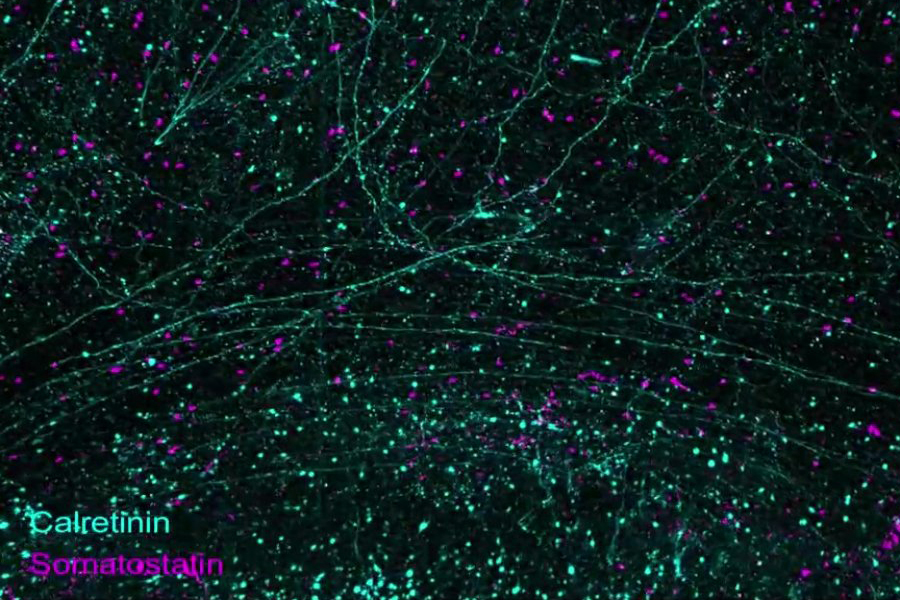
Illustration of precise Megatome slicing preserving anatomical details
Implications for Alzheimer’s Research and Beyond
The UNSLICE system meticulously reassembles these slices at multiple scales, enabling detailed mapping of blood vessels and neural axons. The researchers demonstrated the pipeline’s capabilities through numerous examples, showcasing rich labeling across different cellular structures.
In collaboration with Alzheimer’s researcher Matthew Frosch, Chung’s team began exploring how this pipeline could enhance understanding of Alzheimer’s disease. Their investigations revealed significant neuronal loss in specific areas of diseased tissue compared to controls. Notably, they found that synapse loss correlated with amyloid plaques in certain regions but not others.
While their study involved only two samples and does not draw definitive conclusions about Alzheimer’s disease, it underscores the potential of this technology for deepening our understanding of brain function and pathology.
A Scalable Platform for Organ Research
Importantly, this innovative technology is not limited to brain tissues alone; it holds promise for advancing research across various organs in the body. The authors conclude that this scalable technology platform could significantly enhance our comprehension of organ functions and disease mechanisms, paving the way for the development of new therapies.